Home
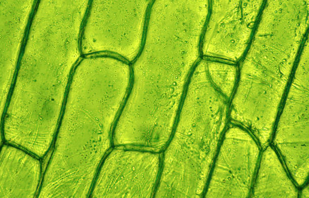
/ Diagram Of Plant Cell As Seen Under Light Microscope - 1 2 Difference Between Plant And Animal Cells Cells As The Basic Units Of Life Siyavula - The cell wall provides and maintains the shape of these cells and serves as a protective barrier.
Diagram Of Plant Cell As Seen Under Light Microscope - 1 2 Difference Between Plant And Animal Cells Cells As The Basic Units Of Life Siyavula - The cell wall provides and maintains the shape of these cells and serves as a protective barrier.
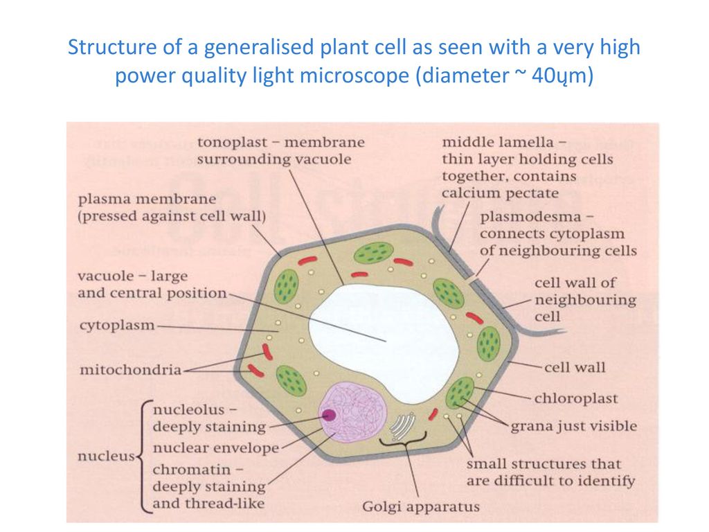
Diagram Of Plant Cell As Seen Under Light Microscope - 1 2 Difference Between Plant And Animal Cells Cells As The Basic Units Of Life Siyavula - The cell wall provides and maintains the shape of these cells and serves as a protective barrier.. Observation of euglena under more powerful electron microscopes have revealed the presence of an ornamented pellicle under the plasma membrane. It is the standard microscope that is used in biology, cellular biology, and microbiological laboratory studies. (a) cell body (b) axon (c) light and dark bands (d) dendrites. Plant cells have a rigid, protective cell wall made up of polysaccharides. The cell membrane (also known as the plasma membrane (pm) or cytoplasmic membrane, and historically referred to as the plasmalemma) is a biological membrane that separates the interior of all cells from the outside environment (the extracellular space) which protects the cell from its environment.
Mar 30, 2017 · a medical student had brought him the ejaculate of a gonorrhea patient to study under his microscope. Preparing a wet mount of a specimen is the technique typically used to view plant and animal cells using a microscope.this page provides step by step instructions on slide preparation as well as videos at the bottom of page. Electron microscopy gives a much higher resolution showing greatly detailed cell structure. (a) cell body (b) axon (c) light and dark bands (d) dendrites. Observation of euglena under more powerful electron microscopes have revealed the presence of an ornamented pellicle under the plasma membrane.
What Is A Diagram Of A Plant And Animal Cell Under An Electron Microscope Quora from qph.fs.quoracdn.net Jun 05, 2021 · brightfield light microscope (compound light microscope) this is the most basic optical microscope used in microbiology laboratories which produces a dark image against a bright background. Mar 30, 2017 · a medical student had brought him the ejaculate of a gonorrhea patient to study under his microscope. Electron microscopy gives a much higher resolution showing greatly detailed cell structure. Fluid collects in the plant cell vacuole and pushes out against the cell wall. Leeuwenhoek obliged, discovered tiny tailed animals, and went on to find the same wriggling. Preparing a wet mount of a specimen is the technique typically used to view plant and animal cells using a microscope.this page provides step by step instructions on slide preparation as well as videos at the bottom of page. In higher plant cells, that polysaccharide is usually cellulose. (a) cell body (b) axon (c) light and dark bands (d) dendrites.
(a) cell body (b) axon (c) light and dark bands (d) dendrites.
May 15, 2021 · brightfield microscope is also known as the compound light microscope. Jun 05, 2021 · brightfield light microscope (compound light microscope) this is the most basic optical microscope used in microbiology laboratories which produces a dark image against a bright background. Plant cells have a rigid, protective cell wall made up of polysaccharides. Unlike most plant cells, this species do not have a cell wall. Pick the odd one out of the following: Preparing a wet mount of a specimen is the technique typically used to view plant and animal cells using a microscope.this page provides step by step instructions on slide preparation as well as videos at the bottom of page. It is an optical microscope that uses light rays to produce a dark image against a bright background. Observation of euglena under more powerful electron microscopes have revealed the presence of an ornamented pellicle under the plasma membrane. Leeuwenhoek obliged, discovered tiny tailed animals, and went on to find the same wriggling. The organelles of the organism and its cytoplasm are therefore bound by a plasma membrane that allows for easier movement. Mar 30, 2017 · a medical student had brought him the ejaculate of a gonorrhea patient to study under his microscope. It is the standard microscope that is used in biology, cellular biology, and microbiological laboratory studies. Made up of two lenses, it is widely used to view plant and animal cell organelles including some parasites such as paramecium after staining with basic stains.
Jun 05, 2021 · brightfield light microscope (compound light microscope) this is the most basic optical microscope used in microbiology laboratories which produces a dark image against a bright background. Mar 30, 2017 · a medical student had brought him the ejaculate of a gonorrhea patient to study under his microscope. Observation of euglena under more powerful electron microscopes have revealed the presence of an ornamented pellicle under the plasma membrane. Pick the odd one out of the following: Fluid collects in the plant cell vacuole and pushes out against the cell wall.
72 221 Plant Cell Stock Photos Pictures Royalty Free Images Istock from media.istockphoto.com Plant cells have a rigid, protective cell wall made up of polysaccharides. Leeuwenhoek obliged, discovered tiny tailed animals, and went on to find the same wriggling. (a) cell body (b) axon (c) light and dark bands (d) dendrites. Observation of euglena under more powerful electron microscopes have revealed the presence of an ornamented pellicle under the plasma membrane. The cells appear elongated tapering at ends as observed under a microscope. Electron microscopy gives a much higher resolution showing greatly detailed cell structure. Unlike most plant cells, this species do not have a cell wall. Cell wall (plant cells only):
Plant cells have a rigid, protective cell wall made up of polysaccharides.
The organelles of the organism and its cytoplasm are therefore bound by a plasma membrane that allows for easier movement. Made up of two lenses, it is widely used to view plant and animal cell organelles including some parasites such as paramecium after staining with basic stains. Unlike most plant cells, this species do not have a cell wall. It is the standard microscope that is used in biology, cellular biology, and microbiological laboratory studies. The cells appear elongated tapering at ends as observed under a microscope. Preparing a wet mount of a specimen is the technique typically used to view plant and animal cells using a microscope.this page provides step by step instructions on slide preparation as well as videos at the bottom of page. Students observed the following tissues under the microscope. Jun 05, 2021 · brightfield light microscope (compound light microscope) this is the most basic optical microscope used in microbiology laboratories which produces a dark image against a bright background. Pick the odd one out of the following: (a) cell body (b) axon (c) light and dark bands (d) dendrites. Mar 30, 2017 · a medical student had brought him the ejaculate of a gonorrhea patient to study under his microscope. May 15, 2021 · brightfield microscope is also known as the compound light microscope. Leeuwenhoek obliged, discovered tiny tailed animals, and went on to find the same wriggling.
Made up of two lenses, it is widely used to view plant and animal cell organelles including some parasites such as paramecium after staining with basic stains. May 15, 2021 · brightfield microscope is also known as the compound light microscope. Fluid collects in the plant cell vacuole and pushes out against the cell wall. The organelles of the organism and its cytoplasm are therefore bound by a plasma membrane that allows for easier movement. Electron microscopy gives a much higher resolution showing greatly detailed cell structure.
Btec Nqf L3 Applied Science Unit 1 Ppt Download from slideplayer.com The cell wall provides and maintains the shape of these cells and serves as a protective barrier. (a) cell body (b) axon (c) light and dark bands (d) dendrites. Preparing a wet mount of a specimen is the technique typically used to view plant and animal cells using a microscope.this page provides step by step instructions on slide preparation as well as videos at the bottom of page. The cells appear elongated tapering at ends as observed under a microscope. Unlike most plant cells, this species do not have a cell wall. Cell wall (plant cells only): It is the standard microscope that is used in biology, cellular biology, and microbiological laboratory studies. In higher plant cells, that polysaccharide is usually cellulose.
Mar 30, 2017 · a medical student had brought him the ejaculate of a gonorrhea patient to study under his microscope.
Plant cells have a rigid, protective cell wall made up of polysaccharides. Fluid collects in the plant cell vacuole and pushes out against the cell wall. The organelles of the organism and its cytoplasm are therefore bound by a plasma membrane that allows for easier movement. The cell membrane (also known as the plasma membrane (pm) or cytoplasmic membrane, and historically referred to as the plasmalemma) is a biological membrane that separates the interior of all cells from the outside environment (the extracellular space) which protects the cell from its environment. Unlike most plant cells, this species do not have a cell wall. The cell wall provides and maintains the shape of these cells and serves as a protective barrier. Observation of euglena under more powerful electron microscopes have revealed the presence of an ornamented pellicle under the plasma membrane. Cell wall (plant cells only): Leeuwenhoek obliged, discovered tiny tailed animals, and went on to find the same wriggling. Students observed the following tissues under the microscope. It is an optical microscope that uses light rays to produce a dark image against a bright background. Preparing a wet mount of a specimen is the technique typically used to view plant and animal cells using a microscope.this page provides step by step instructions on slide preparation as well as videos at the bottom of page. The cells appear elongated tapering at ends as observed under a microscope.
Share :
Post a Comment
for "Diagram Of Plant Cell As Seen Under Light Microscope - 1 2 Difference Between Plant And Animal Cells Cells As The Basic Units Of Life Siyavula - The cell wall provides and maintains the shape of these cells and serves as a protective barrier."

Post a Comment for "Diagram Of Plant Cell As Seen Under Light Microscope - 1 2 Difference Between Plant And Animal Cells Cells As The Basic Units Of Life Siyavula - The cell wall provides and maintains the shape of these cells and serves as a protective barrier."